 - Created by PostDICOM.jpg)
Medical imaging today, in its completely digital form, is expected to meet certain quality standards. Images acquired from the patient are stored in a special format, called the DICOM format (Digital Imaging and Communications in Medicine). This imaging format retains the high quality of the image, and it can only be viewed by specialized viewers known as DICOM viewers. Since these images occupy a lot of space, they also require special software for storage and archiving. This software is referred to as PACS (Picture Archiving and Communication System). Once images are acquired from the patient, they are automatically saved onto the PACS in the DICOM format.
Today, the market is flooded with several DICOM viewers and PACS viewers. How do you choose the right one to suit the needs of your healthcare facility? This article lists some questions that you should ask yourself when looking for the right image viewer.
Most DICOM image viewers today actually need to be ‘PACS viewers’, which means that they should be capable of retrieving an image file from your PACS server—viewing and editing it, and storing it back in the PACS when needed. If your DICOM viewer has an inbuilt mini-PACS, this should not be a problem. However, if you are using a viewer-only software, you must ensure that it is compatible with the PACS server that your healthcare facility uses.
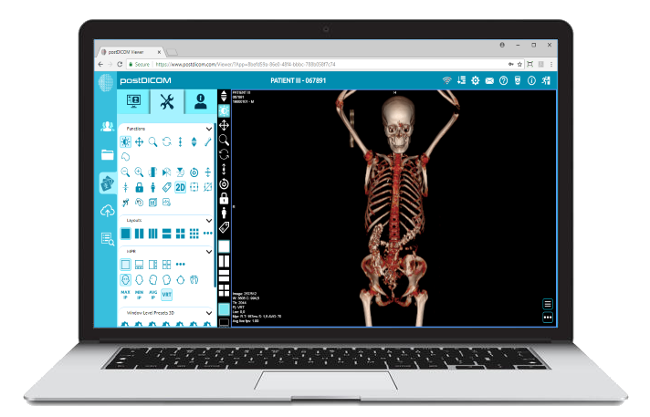
This is no longer an issue, however, if either your DICOM viewer and PACS are cloud-based. Since cloud-based software is essentially hosted on the internet, compatibility is irrelevant. PostDICOM’s PACS cloud viewer, for instance, is accessible through the internet by using a web browser. You can use this to access files from any PACS server. PostDICOM also has a cloud-based PACS system, which you can use to store and retrieve DICOM imaging files.
Using a DICOM viewer, even if it is PACS compatible, would be frustrating if you have to sift through thousands of patient records to access the image that you need. Therefore, your PACS viewer should allow you to easily access a certain patient’s medical records from the archive. For instance, in PostDICOM’s online DICOM viewer, there is a dedicated ‘patient search’ page, where you can search for a record using the patient’s name, ID, or the image modality used. You can access and view both past and current records using the search function.
 - Created by PostDICOM.jpg)
There are several modes of medical imaging today, including CT, MRI, ultrasound, PET, and nuclear bone scans. Each of these have very specific indications, and one imaging modality cannot replace the other. You should, therefore, check if your PACS viewer is able to support all the imaging modalities that are commonly used by your healthcare facility.
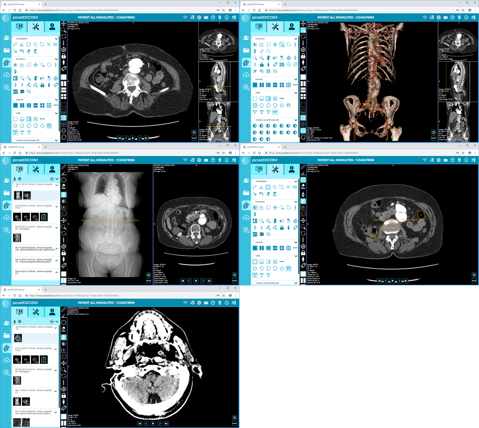
PostDICOM’s imaging viewer supports all medical imaging modalities. It also has a feature called image fusion, which allows you to combine the images acquired by two different modalities into a single image. By doing this, you can get the best out of two different modalities. For instance, the PET-CT fusion allows you to view areas that are metabolically active (a feature of PET scan), and at the same time, allows correct anatomical orientation of the body part (a feature of CT scan).
Most of the PACS viewers on the market may be compatible with only one kind of operating system. If you have Mac OS, you would require a PACS Viewer for Mac, whereas if you have a windows OS, you would need a PACS Windows viewer. Apart from this, if the software is to be installed in your system, you may need a specific amount of space on your hard disk, a minimum RAM speed, and quite possibly an advanced processor. Most applications that are installed on the system are exe or Java-based applications, which may require frequent updates to the latest version.
Today, with the advent of PACS cloud viewers, the need for specific system requirements is removed. Cloud-based DICOM viewers generally use HTML format, which can be accessed using standard web browsers, like Chrome and Firefox. PostDICOM offers one such online viewer, which is an HTML5 PACS viewer, and runs on zero footprint, lossless technology. ‘Zero footprint’ means that the DICOM images do not require any specific loading time. The images can be viewed directly from the location where they are stored and archived. Once the viewing is complete, and the image is stored, there is no trackable history stored on the system. This allows patient data to remain private and confidential.
 - Created by PostDICOM.jpg)
Any image viewer that is used for viewing DICOM files needs to have the following basic functions:
Image enhancement: It is possible to enhance the quality of the image by adjusting its brightness and contrast. Brightness refers to the overall lightness and darkness of the image. When you adjust brightness, every pixel in the image changes to either a lighter shade or a darker shade. Contrast, on the other hand, affects different parts of the image in different ways. When contrast is enhanced, the lighter parts of the image become lighter, while the darker parts of the image become darker. This enhances the sharpness of the image and allows delineation between specific anatomical structures.
Zoom function: This allows us to expand the size of the image (zooming in) and return to the original size (zooming out). The zoom function makes it possible to zero in on the area where an anomaly or disease is suspected, and to examine this part in greater detail.
Pan function: This is designed to go hand in hand with the zoom function. When you zoom in on an image, it may not be possible to view the entire enlarged section in a single frame. The pan function allows you to ‘shift’ the image within the frame, moving it horizontally or vertically.
Rotate and flip functions: In order to gain proper anatomical orientation, it may be necessary to view the image from different angles. Rather than turning your head (or the device!) in all directions, you can use the ‘rotate’ function to turn the image by 90°, 180°, or 270°. The ‘flip’ function allows you to view a mirror image of the selected image frame.
Most DICOM viewers available today feature all the basic functions. In addition to the above mentioned functions, PostDICOM’s online image viewer also allows adjustment of slice thickness in individual images.


|
Cloud PACS and Online DICOM ViewerUpload DICOM images and clinical documents to PostDICOM servers. Store, view, collaborate, and share your medical imaging files. |
Reconstruction modalities—a complete imaging dataset usually consists of a multiple number of images that may be taken in multiple slices but are all in two dimensions. A good PACS viewer should allow you to reconstruct images from these datasets, so that a more thorough interpretation is possible. Some types of reconstruction techniques are as follows:
MPR: MPR stands for Multiplanar reformation. In MPR, data from the complete imaging set is reconstructed to show images in different planes, which were not acquired directly during the imaging process. For instance, a regular CT scan dataset only consists of images that were taken in the axial section. However, using MPR, coronal sections, sagittal sections, and even oblique sections of the body part can also be obtained. A subset of MPR, called curved planar reformation, allows us to obtain curved planes, which may parallel an anatomic structure of interest. For example, aortic vessel analysis can be done by obtaining a curved MPR at a plane that parallels the aorta, and another plane that goes perpendicular to the aorta. This can help assess the diameter of the vessel at any given point.
3D volume rendering: 3D volume rendering takes information from the entire dataset and reconstructs it into a single three-dimensional image. Each individual voxel of the dataset is taken separately, and its contribution is calculated to create a composite image. The advantage of volume rendering over other techniques, like MPR, is that it does not distort anatomical structures during the reconstruction process. Since it essentially combines information from all the images in a dataset, it provides a quick ’overview’ of the entire dataset. Just by glancing at the 3D reconstructed image, it may be possible to identify fractures and pathological lesions. The extent of these anomalies may then be studied in greater detail by going through specific images of the dataset.
Surface rendering: This is also known as Surface Shaded Display, and is meant specifically for delineating the surface of an object in three dimensions. For this, the surface of the area of interest is first ‘separated’ from the underlying structures, using a technique known as segmentation. The surface-defining data is then maintained, while other data are mostly discarded.
Intensity Projections: In Intensity Projections, only images that belong to a specific intensity (that is, within a selected range of Hounsfield Units) are selected. In Maximum Intensity Projection (MIP), for instance, only the objects with highest attenuation values are displayed. This is useful when the area of interest is represented by the brightest objects in the image. For example, MIP is useful to delineate blood vessels in CT and MR angiography, where contrast material is used. Average Intensity projection (AIP) represents average attenuation values, and is primarily used to thicken portions of images that are acquired through MPR. Minimum Intensity Projections (MinIP) display images with lowest attenuation values, and are useful in delineating hypodense areas. For instance, air trapping in the lungs or small airway disease may be detected using MinIP projections.
Obviously, more advanced features and analysis options would increase the utility of your PACS viewer. You would need to choose a PACS viewer keeping in mind which of the above features you are most likely to use. PostDICOM’s online imaging viewer allows MPR, volume and surface rendering, as well as all the intensity projections. PostDICOM also allows calculation of Standard Uptake Values (SUVs) from specialized imaging techniques, such as the PET scan.
 - Created by PostDICOM.jpg)
In your healthcare facility, you may often find it necessary to share images with other physicians for consultation or referral. You would also need to provide patients with copies of their healthcare records. While burning CDs or external disks and transporting them is an effective option, it is time consuming, and not very secure. The recipient would also require a separate PACS viewer to view the DICOM imaging files from the CD or USB drive.
With a cloud-based PACS web viewer, images can be sent securely via email, or can be directly accessed from the PACS website using a single user ID and password. PostDICOM has a cloud-based PACS system that offers its users access from any geographical location, through any device that can access the internet.
DICOM viewing software comes in various price ranges, from zero cost to costing hundreds of dollars. Very often, you need to balance the advanced features of the software against your healthcare facility’s budget. Several DICOM viewers available on the market today offer free versions as well as paid versions. The free versions usually have a free trial version for a short time period or allow only a limited number of patient records to be viewed. Some free versions lack the more advanced features, such as MPR, volume rendering, and surface rendering. You may be able to find and use a PACS viewer free of cost that seems to have all the features, but it may not have the necessary clearances required to facilitate medical diagnosis.
PostDICOM, offers a free to try PACS web viewer and contains all the advanced features that are offered by top viewers on the market. You can start cloud-based PACS storage your free trial now. Cloud storage can be expanded by paying nominal subscription costs on a monthly basis. To take full advantage of these benefits and to experience the features of advanced imaging technology, sign up here.


|
Cloud PACS and Online DICOM ViewerUpload DICOM images and clinical documents to PostDICOM servers. Store, view, collaborate, and share your medical imaging files. |