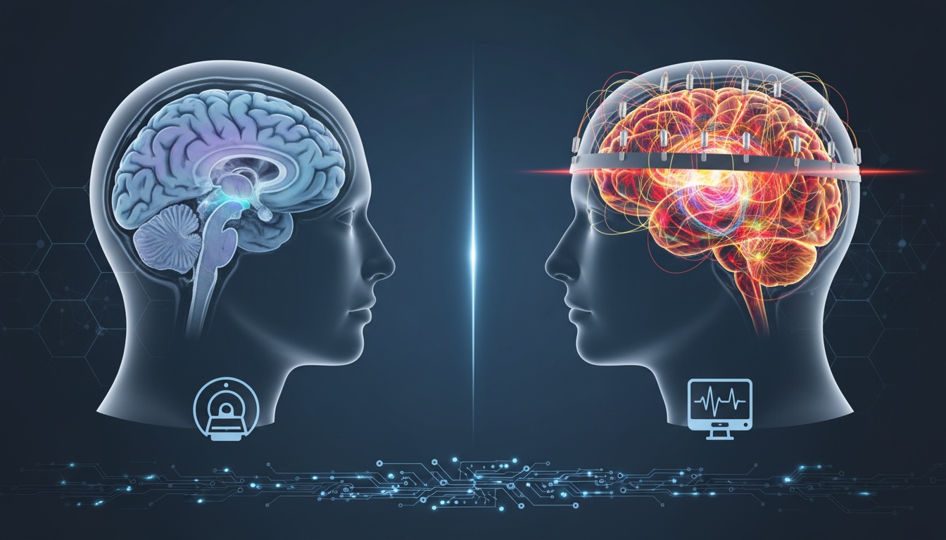
In today's rapidly evolving medical landscape, diagnostic imaging remains a cornerstone of healthcare, offering critical insights into patients' conditions. Magnetic Resonance Imaging (MRI) has long been the gold standard, providing detailed views of the body's structures.
Yet, the Electroencephalogram (EEG) steps into the spotlight when understanding brain function in real time. The EEG's unique ability to track ongoing brain activity offers a dynamic perspective that MRI scans can't capture.
In this blog post, we'll explore the potential of EEGs, their unique capabilities, and specific medical scenarios where they provide insights beyond the reach of MRIs. Join us as we delve into the fascinating world of EEGs and unlock their power in medical diagnostics.
Magnetic Resonance Imaging (MRI) and Electroencephalogram (EEG) are two fundamental diagnostic tools in modern medicine, each with unique capabilities.
MRI is an imaging technique that uses powerful magnets and radio waves to create detailed images of the body's internal structures. It excels in illustrating physical attributes - brain anatomy, soft tissues, and other organs- often used to detect structural abnormalities or damage.
On the other hand, EEG is a neurological test that measures and records the brain's electrical activity. While it may not provide detailed structural images like MRIs, EEGs capture the brain's physiological function in real time.
This includes tracking neural communication, detecting abnormalities in brain waves, and monitoring changes over time, offering unique insights that MRIs cannot provide.
Electroencephalograms (EEGs) have unique capabilities that make them invaluable in neurology and psychiatry. Here's a closer look at how EEGs work and why they are so important:
Unlike other imaging technologies, EEGs can capture the brain's electrical activity as it happens. This allows healthcare providers to monitor brain wave patterns in real time, giving them immediate feedback about changes in brain activityThis is particularly useful in conditions that can cause sudden changes in brain activity, such as epilepsy, as it can capture the exact moment when abnormal brain activity occurs.
Another strength of EEGs is their superior temporal resolution. This means that they can capture changes in brain activity that occur in fractions of a second.
In comparison, MRIs, even functional MRIs (fMRI) that measure brain activity, cannot match the temporal resolution of EEGs. This makes EEGs particularly useful in studying neurological events that happen quickly, such as seizures or certain sleep disorders.
EEGs are non-invasive and can be performed quickly, making them suitable for various clinical situations. For patients who may not be able to undergo an MRI due to certain contraindications (e.g., implanted metallic devices), an EEG can offer an alternative method of investigating brain function.
EEGs measure the brain's electrical activity, essentially communication between neurons. This allows healthcare providers to study how different parts of the brain communicate with each other and detect disruptions in these communications.
This capability can be invaluable in diagnosing and managing disorders that affect neural communication, such as autism and ADHD.
Though MRIs are powerful diagnostic tools, there are several specific medical scenarios where EEGs can provide more nuanced and actionable insights:
In conditions such as epilepsy, an EEG is often the go-to diagnostic tool. While MRIs can identify structural changes or abnormalities that might cause seizures, EEGs are used to record the brain's electrical activity during a seizure.
This allows doctors to classify the seizure type and identify its focus or origin in the brain, which is crucial for effective treatment.
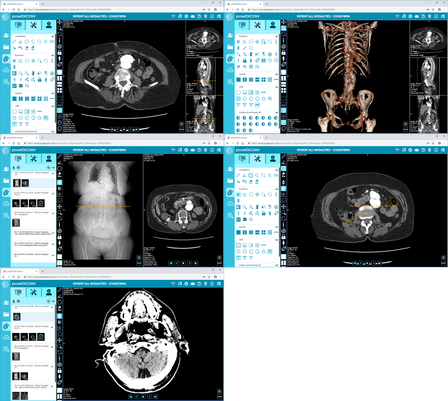
 - Created by PostDICOM.jpg)
Many sleep disorders, including sleep apnea and insomnia, have distinct patterns on EEG.
In polysomnography, a type of sleep study, EEG is used along with other monitoring techniques to observe and record the patient's brain waves, oxygen levels in the blood, heart rate, breathing, and eye and leg movements during sleep. This data can't be captured through MRI, making EEG indispensable in sleep medicine.
Encephalopathies, or diseases that affect the brain's function or structure can often be detected with EEG. Conditions like hepatic encephalopathy or metabolic encephalopathy can produce distinctive EEG patterns even when MRI images appear normal. Thus, EEG can be a valuable tool for diagnosing and managing such conditions.
Certain neurodevelopmental disorders like autism, ADHD, and learning disabilities can show specific EEG patterns. While these disorders can't be diagnosed with EEG alone, EEG can provide supportive evidence and help monitor the effect of treatments on brain activity.
During surgeries that risk affecting brain function, real-time EEG monitoring can alert surgeons to potential issues, such as insufficient blood flow to the brain. This is a critical function that MRI cannot provide.
While MRIs and EEGs each have unique strengths and capabilities, using them together can offer a more comprehensive understanding of the patient's condition. Here's how these two powerful diagnostic tools can complement each other:
MRIs provide exceptional details about the brain's structure, identifying anomalies such as tumors, strokes, or brain injuries.
On the other hand, EEGs illustrate the brain's physiological function. Clinicians can link structural abnormalities with functional ones by using them in conjunction, painting a complete picture of the patient's condition.
MRIs can indicate potential problem areas in the brain's structure but can't specify the type of functional disruption.
EEGs can supplement this information by demonstrating how those structural changes impact the brain's electrical activity. This added layer of detail can refine diagnosis and guide more precise treatment plans.
MRIs can show changes in the brain's structure throughout treatment, such as tumor size reduction. Concurrently, EEGs can track changes in the brain's electrical activity, providing insights into how the brain's function responds to the treatment.
This dual monitoring can help assess the effectiveness of the treatment and adjust it as necessary.
In research contexts, combining EEGs and MRIs can aid in studying brain disorders and developing new treatments.
For instance, simultaneous EEG-fMRI recording is a technique used in neuroscience research to obtain high temporal resolution data from EEG with the spatial resolution of fMRI, giving us a deeper understanding of the brain's workings.
As medical technology continues to advance, we can expect both EEG and MRI technologies to evolve and offer even greater insights into healthcare:
Innovations in EEG technology are promising. For instance, newer devices are becoming more portable and user-friendly, allowing for easier and more widespread use.
Wearable EEG technology could allow long-term, ambulatory monitoring, opening up new possibilities in managing conditions such as epilepsy. Advancements in signal processing algorithms and machine learning enable more accurate EEG data interpretation, improving diagnostic capabilities.
MRI technology advances, with higher magnetic field strengths allowing for even more detailed images. Functional MRIs (fMRIs) and Diffusion Tensor Imaging (DTI), which can provide information about brain activity and white matter integrity, are becoming more commonplace.
There is ongoing research to reduce the noise and the examination time, enhancing patient comfort and compliance.
The future may hold more integrated approaches to combine EEG and MRI data. Sophisticated analytical software could merge structural data from MRIs with functional data from EEGs, offering a holistic view of brain health.
This integration could revolutionize the diagnosis and treatment of many neurological conditions.
Both EEG and MRI are poised to play significant roles in personalized medicine. By providing detailed information about a patient's unique brain structure and function, these tools can help tailor treatments to individual needs, improving efficacy and reducing side effects.
AI and Machine Learning: Artificial intelligence and machine learning are starting to be utilized in analyzing EEG and MRI data, potentially enabling faster, more accurate diagnoses and personalized treatment plans.
In the diagnostic imaging landscape, MRI and EEG hold distinct, invaluable roles. While MRI gives us unparalleled views of the brain's structure, EEG unlocks the dynamic realm of real-time brain function.
They can offer a comprehensive understanding of brain health when used in concert. As technology advances, we can anticipate even greater integration of these tools, paving the way for more precise diagnoses and personalized treatments.
Harnessing the power of EEGs alongside MRIs will continue revolutionizing neurological care, ultimately leading to better patient outcomes in the ever-evolving medical landscape.


|
Cloud PACS and Online DICOM ViewerUpload DICOM images and clinical documents to PostDICOM servers. Store, view, collaborate, and share your medical imaging files. |