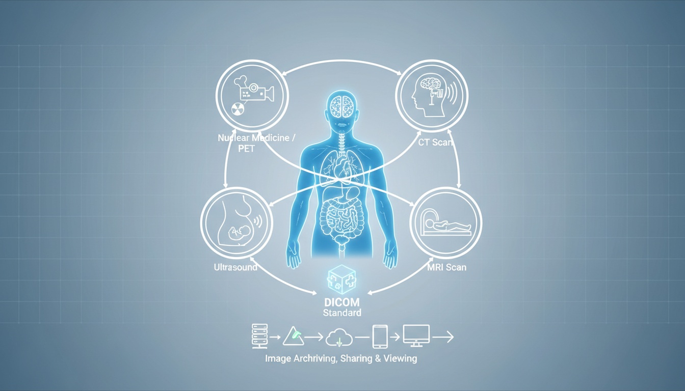
With modern technology in the medical field, doctors are able to diagnose and treat patients without any dangerous side effects. Medical imaging is considered to be one of the best means to achieve that aim, being able to observe what’s happening inside the body without the requirement for surgery or other invasive measures. Medical imaging can be defined as a technique of developing visual representations of areas inside the human body to diagnose health issues and accordingly monitor treatment.
The procedure has had a great impact on public health. Being one of the most powerful resources available for the patients, medical imaging can be used for both therapeutic and diagnostic purposes.
There are numerous kinds of medical imaging, and more means are being developed as technology advances. All kinds operate diversely to develop images of what’s occurring inside the body, so read on to know more.
This particular imaging procedure involves magnetic fields and radio waves to look at the organs and other structures in the human body. The process needs an MRI scanner, which is a huge tube that contains a massive circular magnet. This magnet creates a magnetic field that aligns the protons of hydrogen atoms in the body. The protons are then exposed to radio waves, causing the protons to rotate. When the radio waves are turned off, the protons relax and realign themselves, emitting radio waves in the recovery process that can be sensed by the machine to develop an image.
This imaging procedure makes use of high-frequency sound waves, that is reflected off tissue to develop images of joints, muscles, organs, and soft tissues. It’s like shining light on the inside of the body, except that the light travels through the skin layers and can only be viewed using electronic sensors. Being one of the most cost-effective forms of medical imaging, Ultrasound has no harmful effects and is also regarded as the safest form of medical imaging with a wide range of applications.
This imaging procedure uses electromagnetic radiation to take images of the inside of the body. The most popular and common form of radiography is x-ray. For this imaging procedure, an x-ray machine beams high-energy waves onto the body. The soft tissues, like organs and skin, do not absorb these waves, whereas hard tissues like bones do absorb such waves. The machine transfers the x-ray results onto a film, indicating the body parts that absorbed the waves in white and leaving the unabsorbed material in black.
These are a form of X-ray that develops 3D pictures for diagnosis. Also known as Computed Axial Tomography (CAT), it uses X-rays to develop cross-sectional images of the human body. The scanner has a huge circular opening for the patient to lie on a motorized table. The detector and X-ray then rotate around the patient developing a narrow ‘fan-shaped’ beam of x-rays that passes through a section of the patient’s body to develop an image. CT scans offer greater clarity than conventional x-rays with more precise images of the bones, blood vessels, internal organs, and soft tissue within the body. In most cases, the use of CT scans prevents the requirement for exploratory surgery.
Each procedure is used in diverse situations. For instance, MRI scanners are used to take images of the brain and other internal tissues, specifically when high-resolution images are required. Radiography is used where there is a requirement of images of bone structures to search for breakages. An ultrasound is used to look at fetuses in the womb and takes images of the internal organs when high resolution is not essential.
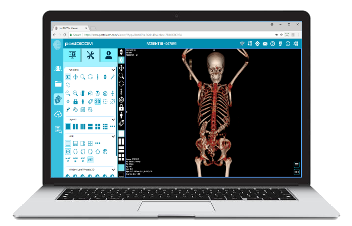
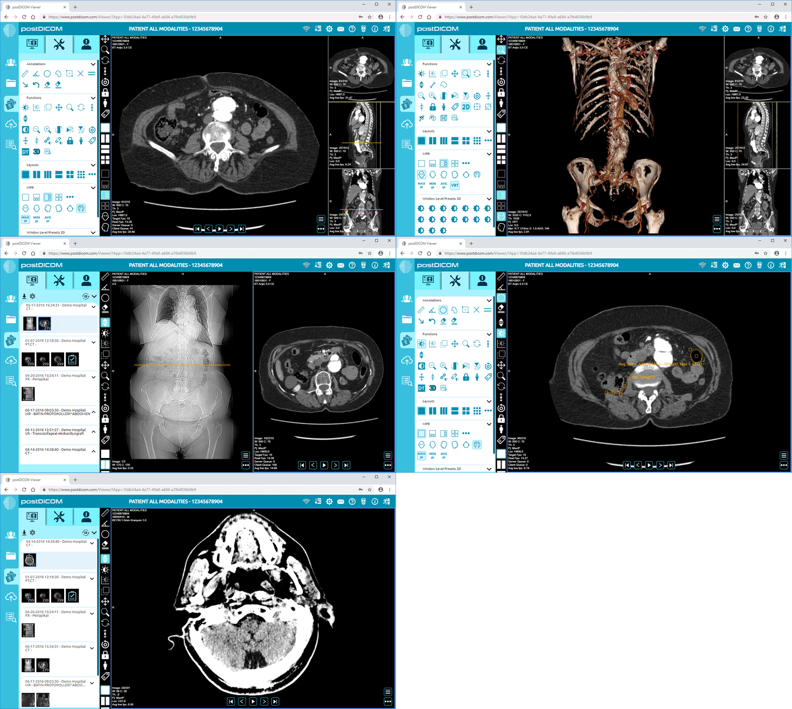
PostDICOM offers two advanced cloud-based medical imaging solutions that make medical image sharing convenient and easy. Once you register with us, you’ll be able to try the viewer free of charge.
We offer a state-of-the-art cloud-based PACS solution, which can be expanded up to 10 TB at nominal costs. There is high security on the services to maintain patient confidentiality and privacy.


|
Cloud PACS and Online DICOM ViewerUpload DICOM images and clinical documents to PostDICOM servers. Store, view, collaborate, and share your medical imaging files. |