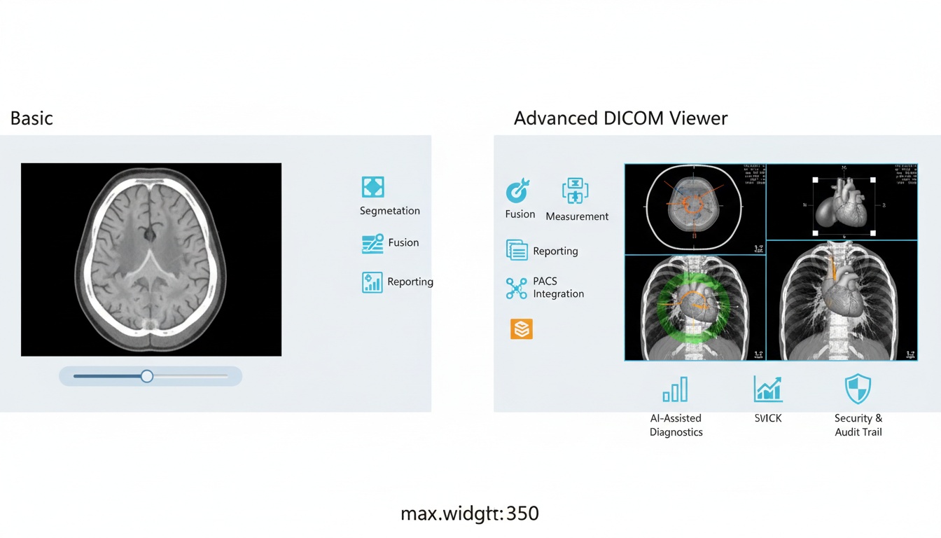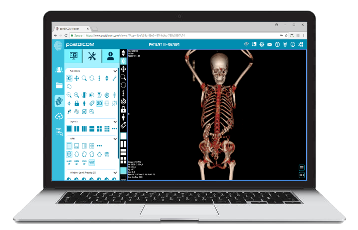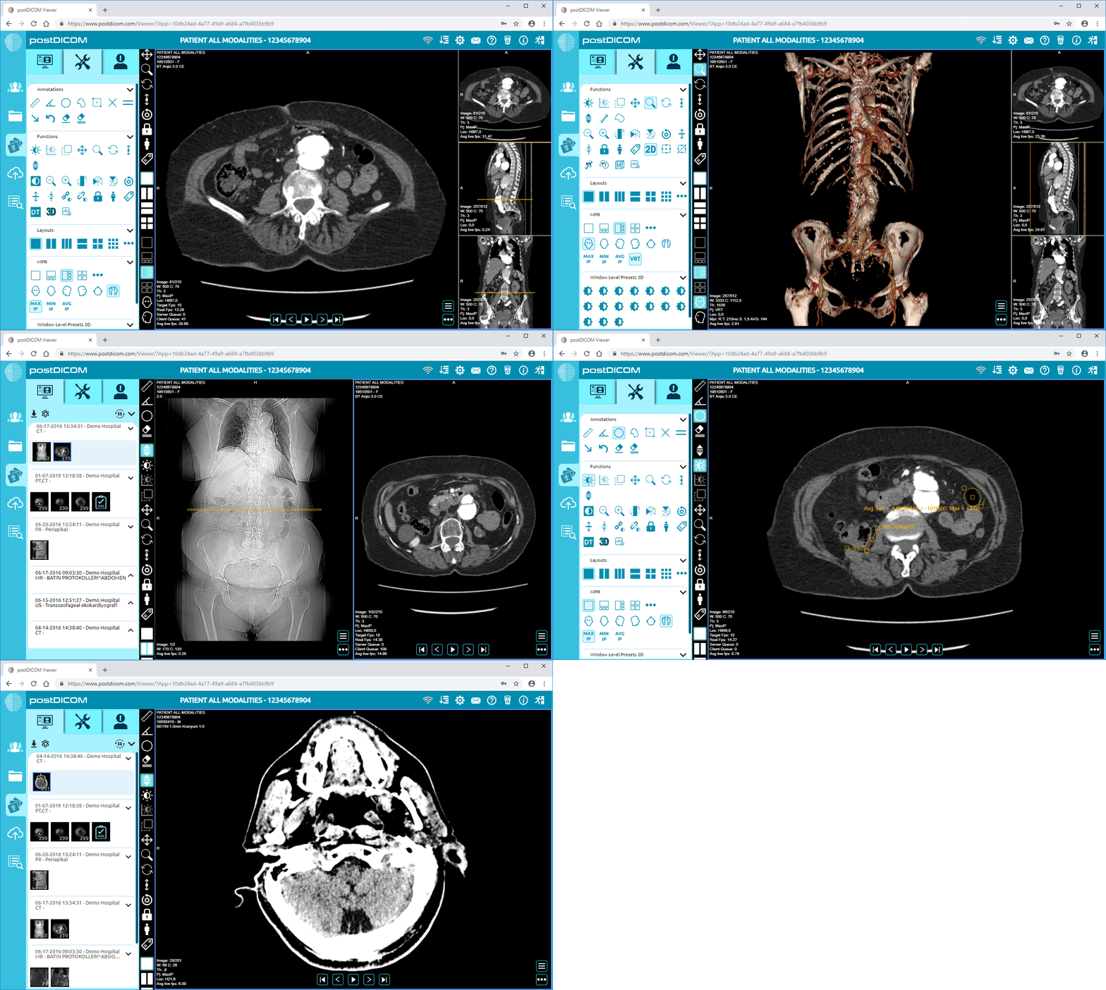
Selecting a suitable DICOM viewer is crucial for any medical or research facility. These software solutions are the gateway to the wealth of information contained in medical images.
The suitable DICOM viewer can streamline workflows, enhance diagnostic capabilities, and, most importantly, contribute to improved patient care. But with the range of available options, how do you know which features are essential for your needs?
It's essential to look beyond basic image-viewing capabilities. Consider factors like user interface, advanced visualization tools, integration with other systems, and security protocols. These features can significantly impact your patient data's efficiency, accuracy, and overall security.
This comprehensive buyer's guide will explore the top features you should prioritize when choosing your next DICOM viewer. We'll provide insights to help you make an informed decision that aligns with your facility's requirements and goals.
In fast-paced healthcare facilities, medical software's usability can significantly impact medical staff effectiveness and efficiency. A DICOM viewer with an intuitive user interface is essential for radiologists and all medical personnel interacting with patient imaging.
Here are the Top 25 Best Free DICOM Viewers for your business!
Such an interface ensures that users can effortlessly navigate the software, regardless of their technical skills or familiarity with digital tools. This ease of use is crucial in high-stress situations where quick access to patient data and images can directly influence patient outcomes.
A well-designed user interface goes beyond simple aesthetics; it creates an efficient workflow that reduces the steps needed to perform common tasks. For instance, features like drag-and-drop functionality, easy-to-navigate menus, and customizable toolbars allow users to access frequently used functions quickly, saving time and reducing the cognitive load on staff. This streamlined interaction minimizes the chances of errors and enhances the focus on patient care rather than on navigating the software.
A user-friendly DICOM viewer's simplicity also significantly reduces staff training time. When the software is intuitive, new users can familiarize themselves with essential functions more quickly and become proficient without extensive training.
This is particularly beneficial in high-turnover environments and where time resources are scarce. Facilities can allocate their training resources to other critical areas, enhancing operational efficiency.
Ease of use in software design supports current staff and plays a pivotal role in the broader adoption of the technology within a facility. When medical personnel interact positively with the DICOM viewer, the likelihood of its acceptance and continued use increases.
This user satisfaction translates into better compliance with imaging protocols and more consistent, reliable usage of imaging software, directly impacting patient care quality.
Image quality and clarity in medical imaging are paramount for accurate diagnosis and effective patient care. Advanced image processing features in DICOM viewers, such as contrast adjustment, zoom, and 3D rendering, are essential tools that empower healthcare professionals to view and analyze medical images with greater precision.
These capabilities improve the visibility of subtle anatomical details and significantly enhance the diagnostic process.
Contrast adjustment is a critical feature in any DICOM viewer. It allows radiologists and other medical professionals to modify the lightness and darkness of an image, thereby highlighting specific features or areas of interest.
This is particularly crucial in detecting fine details in soft tissues or in visualizing slight variations in density that could indicate the presence of abnormalities such as tumors or fractures.
Zoom functionality, meanwhile, plays a vital role in examining specific areas of an image more closely. By enabling users to focus on minute details without losing resolution, zoom can help identify early signs of disease that might otherwise go unnoticed. This capability is indispensable in fields such as oncology, where early detection can significantly affect the outcome of treatments.
3D rendering takes two-dimensional images and reconstructs them into three-dimensional models, providing a holistic view of anatomical structures that can be crucial for complex diagnoses and surgical planning.
This feature allows doctors to navigate around, through, and within the 3D model, offering perspectives that are impossible to achieve with traditional 2D images. For instance, in orthopedics, 3D models of bone structures can help surgeons plan joint replacements or reconstructive surgeries with greater accuracy.
The clarity and detail provided by these advanced image-processing tools are invaluable in medical settings. Enhanced image quality ensures that clinicians can make more informed and confident decisions.
For example, more explicit images reduce the likelihood of misdiagnosis and enable more precise targeting during biopsy procedures or radiation therapy, directly impacting patient safety and treatment efficacy.
In the healthcare industry, protecting patient information is a matter of ethical practice and a legal requirement.
Modern DICOM viewers are equipped with advanced security features that ensure compliance with stringent healthcare regulations, such as the Health Insurance Portability and Accountability Act (HIPAA) in the United States. These features are crucial for maintaining the integrity and confidentiality of patient data.
Encryption is one of the fundamental security measures in any DICOM viewer. It ensures that all medical images and associated data are encrypted in transit and at rest. This means that any data sent over networks or stored on servers is converted into a secure format that is nearly impossible to decrypt without authorized access.
Encryption protects against unauthorized data breaches and ensures that patient information remains confidential, thus upholding patients' trust in healthcare providers.
Secure access controls are critical to prevent unauthorized access to sensitive medical data. DICOM viewers typically feature robust authentication mechanisms, including strong password policies, multi-factor authentication, and role-based access controls.
These measures ensure that only authorized personnel can access patient images and data based on their roles and responsibilities within the healthcare facility. This selective accessibility helps minimize the risk of accidental or malicious data exposure.
Audit trails are another vital security feature of DICOM viewers. They provide a detailed log of all activities related to patient data, such as who accessed the data, when, and for what purpose. This tracking is crucial for detecting and responding to potential security violations.
Audit trails are also invaluable for regulatory compliance, providing verifiable proof that the facility adheres to guidelines regarding handling and protecting patient information.
Together, these security features form a comprehensive defense mechanism against the increasing threats to data security in the healthcare sector.
DICOM viewers protect patient data and help healthcare facilities comply with relevant regulations by implementing strong encryption, secure access controls, and thorough audit trails. This level of security is essential for building and maintaining trust between patients and providers.
In the interconnected environment of modern healthcare facilities, the ability of different systems to work together seamlessly is not just advantageous—it’s essential.
DICOM viewers are often at the heart of medical imaging workflows, and their ability to integrate with other hospital systems, such as Electronic Health Records (EHRs) and Picture Archiving and Communication Systems (PACS), significantly affects a facility's operational efficiency and the quality of patient care.
When DICOM viewers are fully compatible and integrated with EHRs, all patient data, including images, reports, and other medical records, are centralized and accessible through a single interface. This integration means physicians can access complete patient histories, diagnostic images, and other relevant information without switching between systems.
Such accessibility saves valuable time and reduces the mental load on healthcare providers, allowing them to focus more on patient care rather than navigating through various software systems.
The seamless integration of DICOM viewers with PACS and EHRs significantly reduces the likelihood of errors. Automating the transfer of images and data between systems minimizes the risk of manual entry errors.
For example, when a DICOM viewer is linked directly to an EHR system, the images taken from a PACS are automatically updated in the patient’s electronic health record.
This automatic updating ensures that every healthcare team member has access to the most current and accurate information, leading to more informed decision-making and improved patient outcomes.
Integration also streamlines diagnostic processes by enhancing the flow of information. Radiologists and other specialists can quickly upload their findings and images to a shared system where different departments can easily access and review them.
This capability is particularly crucial in multidisciplinary cases involving several specialists in diagnosis and treatment planning.
Quick and easy access to updated, comprehensive patient information facilitates better communication among caregivers and leads to more coordinated and effective care.
The integration of cloud technology in DICOM viewers has transformed the landscape of medical imaging and diagnostics. Cloud-based DICOM viewers allow healthcare professionals to access medical images and related data from anywhere, anytime, using any compatible device.
This level of accessibility is crucial for the growing field of telemedicine and for healthcare providers who need to make timely decisions in critical care scenarios.
Telemedicine relies heavily on seamlessly and securely sharing medical information across different locations. Cloud-based DICOM viewers facilitate this this by allowing images to be viewed and shared in real time without physically transporting image files.
This capability supports remote diagnostics and consultations and enhances collaborative healthcare by enabling multiple specialists to review and discuss the same medical images simultaneously, regardless of their physical locations. Such collaborations can lead to more comprehensive evaluations and improve patient outcomes.
One of the most significant advantages of using cloud-based DICOM viewers is scalability. Healthcare facilities can adjust their storage needs based on current demand without substantial upfront investments in physical infrastructure.
As a facility grows or the volume of imaging data increases, cloud storage can be easily expanded to accommodate these changes, ensuring the system adapts smoothly to evolving needs.
Another critical benefit of cloud storage in medical imaging is enhanced disaster recovery. Traditional on-premises storage solutions are vulnerable to physical damages, such as those caused by natural disasters or system failures.
Cloud storage provides robust disaster recovery solutions by duplicating data across multiple secure locations, ensuring that data can be quickly restored and accessed even if one storage site is compromised.
This redundancy protects data and ensures that healthcare operations can continue without significant interruptions, a crucial factor in maintaining patient care continuity.
In the complex field of medical diagnostics, it is invaluable to view and analyze images from various modalities within a single DICOM viewer.
Each imaging modality—be it X-ray, MRI, or CT—provides unique insights into the body’s internal structures and functions. X-rays are excellent for examining bone fractures and alignments; MRIs provide detailed images of soft tissues, including the brain and muscles; and CT scans offer a comprehensive overview of the body’s anatomy, including bones, blood vessels, and soft tissues.
Supporting these varied modalities within one viewer allows clinicians to cross-reference and compare images for a more holistic assessment of a patient’s condition.
Multi-modality support in DICOM viewers streamlines the diagnostic process by consolidating all relevant patient images into one accessible platform.
Clinicians no longer need to switch between systems to view images from different sources. This integration significantly speeds up the review process, reduces the risk of overlooking critical information, and enhances workflow efficiency.
For instance, a radiologist examining a patient with a complex joint disorder can simultaneously view MRI images for soft tissue assessment and X-ray images for bone structure analysis, facilitating a faster and more accurate diagnosis.
The capability to support multiple imaging modalities is particularly beneficial in multidisciplinary medical settings where various specialists collaborate on patient care.
For example, combining PET scans with CT scans within the same viewer can provide a more detailed visualization of a tumor’s metabolic activity and anatomical position in oncology.
This integrated view helps oncologists and surgeons plan more effective treatment strategies, such as targeted radiation therapy or surgical interventions, ultimately leading to better patient outcomes.
Multi-modality support also plays a crucial role in personalized medicine, where treatment plans are tailored to each patient's individual characteristics.
By providing a comprehensive picture of a patient’s medical condition through various imaging modalities, DICOM viewers equipped with multi-modality support enable healthcare providers to customize treatments based on detailed, accurate data.
This approach improves the effectiveness of treatments and minimizes potential side effects by avoiding unnecessary procedures.
 - Created by PostDICOM.jpg)
Customization in DICOM viewers is crucial for aligning the software capabilities with a healthcare facility's specific needs. Each facility has unique workflow patterns, specialist requirements, and patient care protocols.
DICOM viewers can be tailored to enhance these specific operational workflows by allowing customization of features, thereby increasing efficiency and productivity.
For instance, a facility specializing in neurology might require advanced neuroimaging tools integrated into its DICOM view. At the same time, a general hospital might focus on tools that provide broader utility across various medical disciplines.
Customization helps streamline daily operations by adapting the software’s interface and functionality to the user’s preferences and frequently performed tasks.
For example, if a radiology department frequently conducts certain types of analyses, having these tools readily accessible on the main dashboard of the DICOM viewer reduces the time spent navigating through menus.
This direct access speeds up the diagnostic process and reduces clinicians' cognitive load, allowing them to focus more on patient care rather than on managing the software.
As healthcare facilities expand, so does the volume of their imaging data and the complexity of their operational needs. Scalability in a DICOM viewer refers to the system’s ability to handle increased workloads and expanded functionalities without compromising performance or requiring a complete system overhaul.
This means that as a hospital grows and evolves—perhaps by adding new departments or specialties—the DICOM viewer can scale up to meet these new demands. Scalable systems are crucial for long-term planning and sustainability, ensuring that the initial technology investment continues to deliver value over time.
A scalable DICOM viewer can accommodate everything from an increase in the number of users as the facility hires more staff to the integration of new imaging modalities and advanced analytical tools as medical technology advances.
This flexibility is especially important in multi-site healthcare organizations, where standardized yet adaptable tools are necessary to ensure consistent quality of care across all locations.
Scalability ensures that new modules or upgrades can be integrated smoothly, allowing facilities to adopt the latest innovations in medical imaging without encountering significant disruptions or compatibility issues.
Remember, investing in a robust DICOM viewer is not just about the software itself – it's about empowering your medical professionals with the tools they need to deliver the best possible patient care. Consider these factors carefully:
Vendor Support: Choose a vendor with a proven track record of excellent customer service and ongoing technical support.
Future-Proofing: Look for a DICOM viewer solution that's adaptable and regularly updated to keep pace with evolving technology and standards.
Trial Periods: Take advantage of free trials or demos whenever possible to get hands-on experience before purchasing.
Choosing the right DICOM viewer can be complex, but the benefits are well worth the effort. By prioritizing the features outlined in this guide, you'll find a solution that optimizes your workflows, advances your diagnostic capabilities, and ultimately enhances the quality of care you provide to your patients.


|
Cloud PACS and Online DICOM ViewerUpload DICOM images and clinical documents to PostDICOM servers. Store, view, collaborate, and share your medical imaging files. |